Inputs → figure
Medical Illustration Generator
Describe an anatomical scene or add a reference image to create an AI illustration draft. Review the anatomy and labels; AI output remains a bitmap.
Checking AI availability…
Example input
Start with a reproducible example
Self-authored, simulated teaching input for exploring this workflow. It is not a user’s research or a public gallery submission.
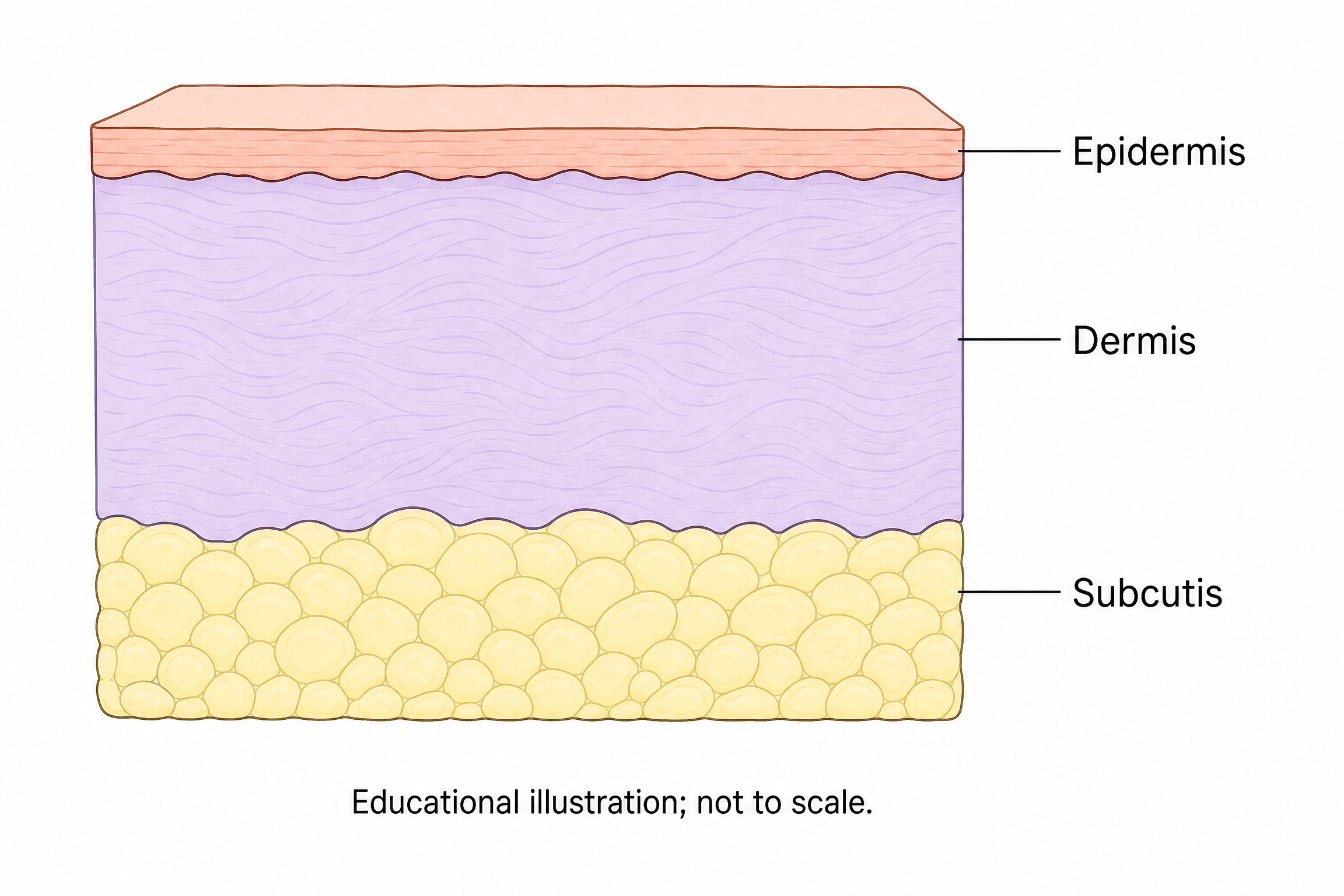
Self-authored teaching cross-section: thin Epidermis above thicker Dermis, then fatty Subcutis; exact three labels and Educational illustration; not to scale. Image 2 2K, 5 provider credits; a simplified teaching draft without expert certification.
Reviewed output · self-authored simulated input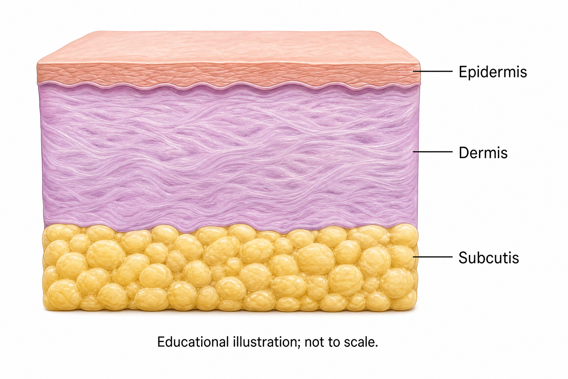
Use the reviewed line-art PNG as reference. Request semi-realistic soft rose/violet/gold, preserving layer order, exact labels and footer. Real workers.dev private-reference upload and Image 2 2K image-to-image output, 5 provider credits.
Reviewed output · self-authored simulated inputRead the example input
{
  "brief": "Create a simplified teaching cross-section of skin on a white background. Show a thin Epidermis at the top, a thicker Dermis directly beneath it, and Subcutis with rounded fat lobules below the Dermis. Keep the three layer boundaries and their top-to-bottom order clear. Use restrained peach, lavender and pale yellow. Include exactly these three labels, each once beside its corresponding layer: \"Epidermis\", \"Dermis\", \"Subcutis\". Add this footer verbatim: \"Educational illustration; not to scale.\" Do not include hair, glands, vessels, nerves, measurements, disease or additional labels.",
  "style": "line-art"
}
Load the skin-layer brief or reference-edit brief to review the supplied input. These are independent AI raster examples; results vary between runs.
How an AI anatomy illustration is created and reviewed
The implemented workspace sends an explicitly confirmed anatomical brief and line-art or semi-realistic style to Image2, fixed at 2K and 3:2 for one raster image. An optional single PNG reference can guide image-to-image revision. The reference must be no larger than 5 MiB, 8192 pixels per side and 24 million pixels total. The current provider estimate is 5 credits per image request; actual reported usage remains visible with the task.
The brief and optional reference are sent to KIE and stored in the shared private test workspace. Reference editing uses a temporary provider upload plus a private copy; it does not guarantee pixel-perfect preservation or anatomical correctness. Labels are pixels inside the raster, with no independent editable annotation layer. Review anatomy, every word and reference drift before use; this is not a diagnostic service, clinical guidance or expert-certified illustration. Base generation and a reference-guided revision have been tested with the teaching example; every new result still requires review.
What this tool does not do
This workspace generates or revises an independent Image2 raster draft. It does not diagnose, provide clinical guidance, certify anatomy or guarantee preservation of a reference. Base generation and reference-guided revision have been tested on the teaching example, without expert certification.
Labeling and accuracy
Requested labels are rendered into the image as pixels. Review each word and anatomical relationship against the brief and reference. Use only material you may send; do not include patient identifiers or confidential clinical material in this shared private test workspace.
Journal and textbook policy
Check the current policies of the intended publication or textbook, including image-source rights, AI disclosure and any restrictions. Choosing a style or reference does not settle those requirements or imply scientific review.
How to use this tool
- Describe only anatomy, view, exact labels and edits you can review; choose line art or semi-realistic style.
- Optionally select one eligible PNG and confirm rights to send it and absence of patient identifiers.
- Review the fixed Image2 2K/3:2 provider estimate, then explicitly generate one image.
- Keep the task ID and check an uncertain result rather than automatically resubmitting.
- Review anatomy, text and edit drift; download the private original PNG only for the current unchanged input.
Specifications and limits
- Input
- Anatomical brief and explicit style; optional one PNG ≤5 MiB, ≤8192px per side and ≤24M pixels
- Styles
- Line-art or semi-realistic directions; Image2 fixed 2K/3:2, estimate 5 provider credits per image
- Output
- Independent raster image; labels are pixels; no native vector or editable annotation overlay
- Review
- User scientific review required; no diagnostic, clinical, anatomical-accuracy or expert-certification guarantee
- Formats
- Private original PNG download; changed or invalid inputs block old-result download
Guest native edits stay in this tab until you explicitly save a confirmed figure. Sign in to keep private works and revision history. Saved native works restore the figure and exports, not the original input form. AI tasks, images and references are private to their owner. Subscriptions and one-time packs are available for AI allowances. Native drawing and exports remain free. Understand export formats.
Frequently asked questions
Can I use a reference image?
Yes. Select one PNG up to 5 MiB, 8192 pixels per side and 24 million pixels total. Confirm you may send it to KIE and the shared private workspace, and that it contains no patient identifiers.
Can I edit words inside the generated image?
You can request a visual revision in the brief using an eligible reference image. The resulting words remain pixels; no independent editable text layer or exact preservation guarantee is provided.
Is the anatomy guaranteed to be accurate?
No. AI can alter relationships, omit structures or render labels incorrectly. Have an appropriate subject-matter expert review the result before factual use; no expert certification is implied.
Is this a diagnostic tool?
No. It creates educational illustration drafts and does not provide diagnosis, clinical guidance or clinical decision support.
Can I download an SVG?
The supported output is an original PNG raster image. There is no native vector SVG or independently editable label overlay.
Will a journal accept the illustration?
Check the intended publication's current AI-image, disclosure and source policies. Generation does not guarantee acceptance or establish third-party usage rights.