
Inputs → figure
Food Digestion Diagram Generator
Create an AI food digestion pathway diagram from supplied organs and labels. Review the mouth-to-anus sequence and download the original PNG.
Checking AI availability…
Example input
Start with a reproducible example
Self-authored, simulated teaching input for exploring this workflow. It is not a user’s research or a public gallery submission.
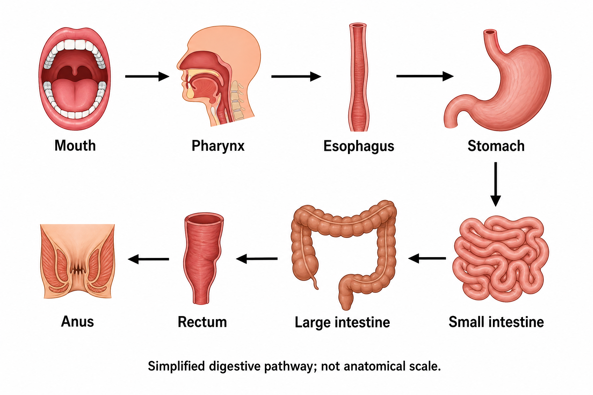
Create a full-color conceptual teaching diagram of the human digestive tract pathway on white, using eight clearly separated organ icons in a two-row snake layout. The top row, left to right, is Mouth, Pharynx, Esophagus, Stomach. A down arrow from Stomach leads to Small intestine at the far right of the bottom row. The bottom row then continues right to left: Small intestine, Large intestine, Rectum, Anus. Connect the eight stages with exactly seven forward arrows, never a closed loop. Put the exact eight organ names beside their corresponding icons. Use readable dark labels and distinct restrained colors. Show only the alimentary pathway; do not route food through a liver or pancreas. Do not add enzymes, durations, nutrient percentages, treatments or measured results. Add the exact footer: Simplified digestive pathway; not anatomical scale.
Reviewed output · self-authored simulated inputRead the example input
{
  "style": "flat-color",
  "brief": "Create a full-color conceptual teaching diagram of the human digestive tract pathway on white, using eight clearly separated organ icons in a two-row snake layout. The top row, left to right, is Mouth, Pharynx, Esophagus, Stomach. A down arrow from Stomach leads to Small intestine at the far right of the bottom row. The bottom row then continues right to left: Small intestine, Large intestine, Rectum, Anus. Connect the eight stages with exactly seven forward arrows, never a closed loop. Put the exact eight organ names beside their corresponding icons. Use readable dark labels and distinct restrained colors. Show only the alimentary pathway; do not route food through a liver or pancreas. Do not add enzymes, durations, nutrient percentages, treatments or measured results. Add the exact footer: Simplified digestive pathway; not anatomical scale."
}
Load the disclosed simulated illustration brief and review every label, connection and scientific detail. AI results vary between runs.
Follow food through the alimentary pathway
The default alimentary pathway diagram follows eight named stages: Mouth, Pharynx, Esophagus, Stomach, Small intestine, Large intestine, Rectum and Anus. It uses separated organ icons and seven forward arrows so you can trace the sequence without mistaking the two-row layout for a closed cycle. Food is not routed through accessory organs such as the liver or pancreas.
Image 2 at 2K creates an independent raster draft from the confirmed brief. Inspect all seven connections, especially the transition from the top row to the lower row. This is a simplified pathway illustration, not an anatomical reconstruction, an enzyme simulation or a calculation of digestion time and nutrient absorption.
Count eight stages and seven forward arrows
Read the organ names in pathway order rather than by page position. Verify the Stomach-to-Small intestine connection crosses between rows only once and that Anus has no return arrow to Mouth.
Distinguish a pathway from anatomical position
Separated icons make the route legible but do not show the organs at anatomical scale or in their actual spatial arrangement. Keep the exact simplified-pathway footer visible when using the image as a teaching aid.
Keep digestive mechanisms within supplied evidence
Do not interpret an organ icon or arrow as a claim about enzyme activity, residence time, disease or nutrient yield. Those details require their own supplied sources and scientific review; the default example makes none of those claims.
Review reference: OpenStax: digestive processes. Check every label, direction and structural assumption against an appropriate subject reference.
How to use this tool
- Describe the subject and the exact relationships to show.
- Choose a style and aspect ratio; optionally add one permitted PNG reference.
- Review the brief and explicitly confirm generation.
- Inspect the result against the supplied scientific details.
- Download the original PNG or revise the brief for a new image.
Specifications and limits
- Route
- Independent Image 2 2K raster illustration; separate from native diagram engines
- Controls
- Scientific brief; line art, flat color or semi-realistic style; supported aspect ratios; optional PNG reference
- Reference
- One PNG up to 5 MiB, 8192 pixels per side and 24 million pixels; only material you may send
- Output
- Original PNG; labels are pixels, without SVG, PPTX or independently editable annotations
- Review
- User scientific review required; no scientific-accuracy or publication-acceptance guarantee
- Scope
- Supplied alimentary pathway; no enzyme simulation or nutrient calculations
Guest native edits stay in this tab until you explicitly save a confirmed figure. Sign in to keep private works and revision history. Saved native works restore the figure and exports, not the original input form. AI tasks, images and references are private to their owner. Subscriptions and one-time packs are available for AI allowances. Native drawing and exports remain free. Understand export formats.
Frequently asked questions
Which organs are included in the default digestive pathway?
The example requests Mouth, Pharynx, Esophagus, Stomach, Small intestine, Large intestine, Rectum and Anus, in that order. These are eight simplified stages with seven forward arrows and an explicit not-anatomical-scale footer.
Why are the liver and pancreas outside the food pathway?
The default example follows the alimentary pathway and excludes the liver and pancreas from that route. If you request accessory organs in another brief, distinguish their relationship to the tract; do not draw food traveling through them.
Does this simulate enzymes or nutrient absorption?
No. The example deliberately omits enzymes, durations and nutrient percentages. The generator produces an illustration from your brief and does not calculate digestive reactions, absorption or physiological measurements.
How should I check the two-row layout?
Start at Mouth and follow each arrow to Anus. Stomach connects downward to Small intestine at the far right of the lower row, which continues right to left. Reject any missing stage, reversed arrow or closed loop.
Can I use my own digestive-system PNG as a reference?
You can attach one eligible PNG you have permission to send to the provider. Specify the intended sequence and exact labels again; reference-guided generation can change organ shapes, labels and connections and requires a fresh review.
Can I edit the digestive labels after downloading?
The original PNG contains labels and arrows as pixels. This route does not export editable anatomical objects, SVG or PPTX shapes. Revise the brief and explicitly generate a new draft to request changes.