
Inputs → figure
Neuron Diagram Generator
Describe a neuron and create an AI teaching diagram with supplied structures and labels. Review branches and signal direction before downloading the original PNG.
Checking AI availability…
Example input
Start with a reproducible example
Self-authored, simulated teaching input for exploring this workflow. It is not a user’s research or a public gallery submission.
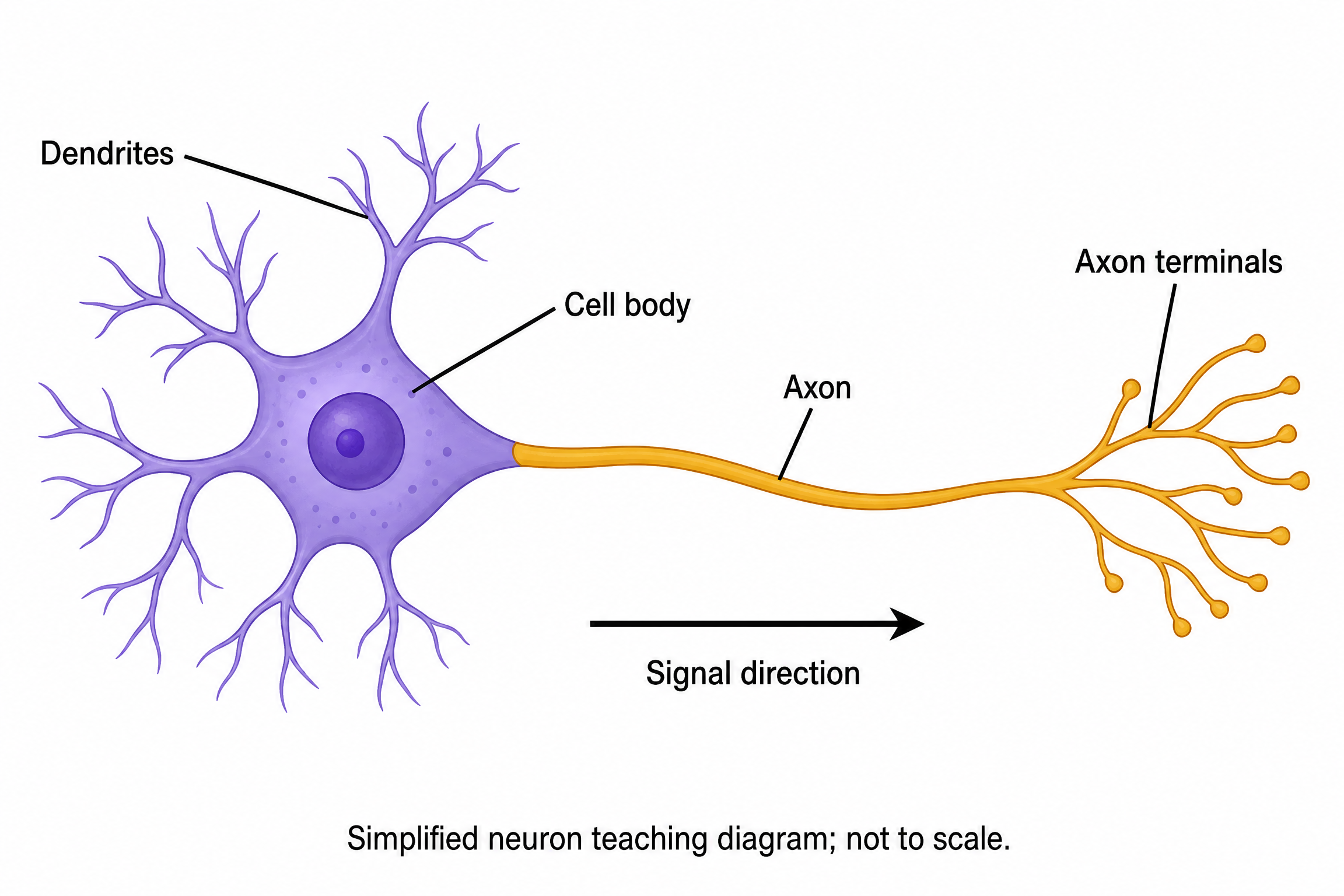
Create a full-color conceptual teaching illustration of one generalized multipolar neuron on white. On the left show branched dendrites connected to a large cell body containing one nucleus. One continuous axon leaves the cell body and runs to branched axon terminals on the right. Use exactly four component labels with leader lines: Dendrites, Cell body, Axon, Axon terminals. Place one clear right-pointing arrow below the axon labeled Signal direction. Do not add myelin, synapses, neurotransmitters, ion channels, measured signals or medical claims. Add the exact footer: Simplified neuron teaching diagram; not to scale.
Reviewed output · self-authored simulated input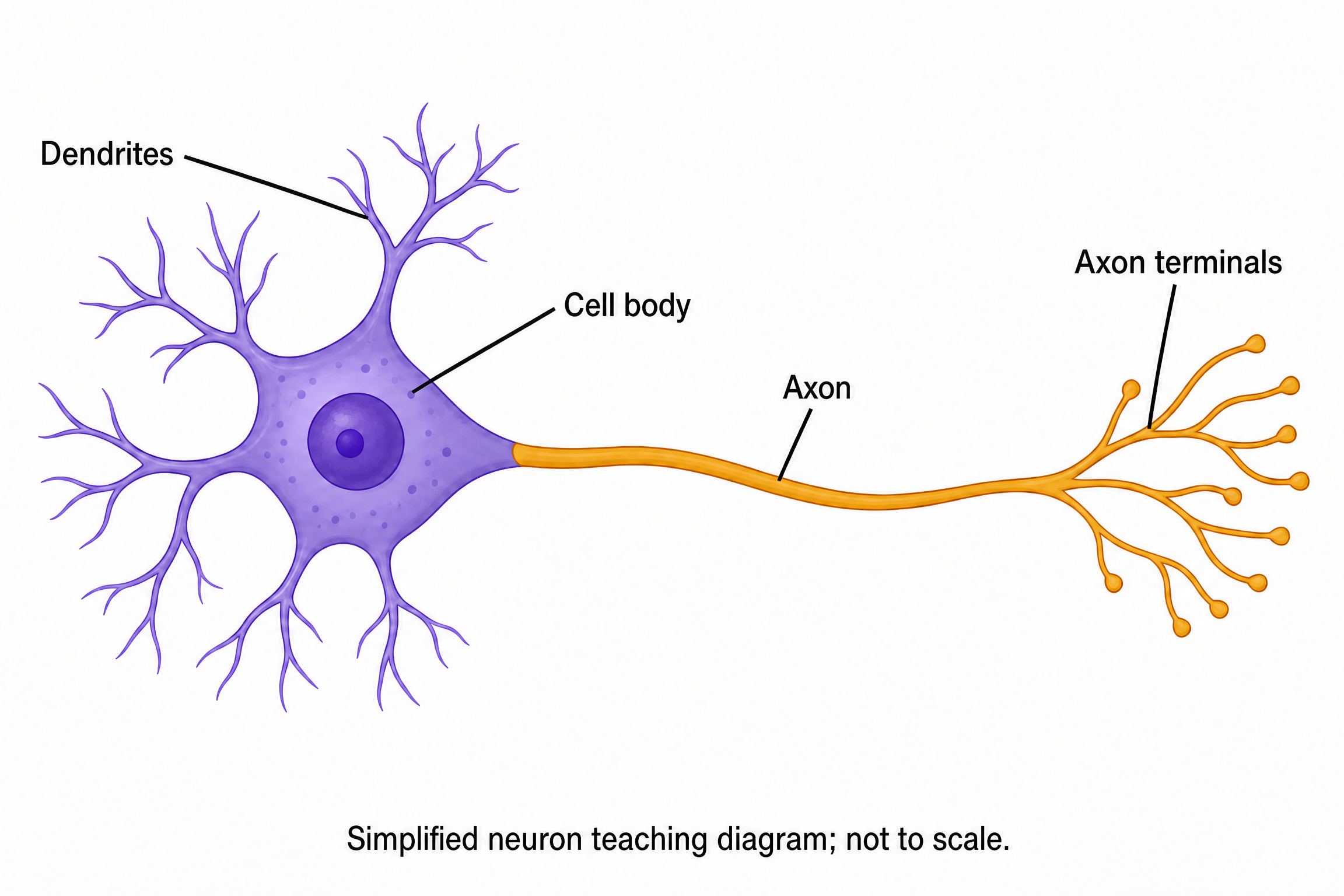
Use a permitted self-authored PNG from the generalized multipolar-neuron example as a reference. Remove only the Signal direction arrow and its label. Preserve the four exact component labels, connected dendrites, cell body, continuous axon, axon terminals and simplified/not-to-scale footer. Add no myelin or synapse.
Reviewed output · self-authored simulated inputRead the example input
{
  "style": "flat-color",
  "brief": "Create a full-color conceptual teaching illustration of one generalized multipolar neuron on white. On the left show branched dendrites connected to a large cell body containing one nucleus. One continuous axon leaves the cell body and runs to branched axon terminals on the right. Use exactly four component labels with leader lines: Dendrites, Cell body, Axon, Axon terminals. Place one clear right-pointing arrow below the axon labeled Signal direction. Do not add myelin, synapses, neurotransmitters, ion channels, measured signals or medical claims. Add the exact footer: Simplified neuron teaching diagram; not to scale."
}
Load the disclosed simulated illustration brief and review every label, connection and scientific detail. AI results vary between runs.
Describe the neuron’s branches and direction
A neuron teaching diagram separates dendrites, the cell body, an axon and axon terminals. The default brief describes one generalized multipolar neuron, with dendrites to the left and a continuous axon reaching terminals to the right. It omits myelin and synaptic detail so the requested component relationships are easier to inspect.
Image 2 at 2K creates an independent raster illustration from the supplied brief. Trace the axon from the cell body to the terminals and inspect the requested signal arrow separately from the label leader lines. This simplified direction cue does not model electrical activity, measure a nerve signal or describe every neuron type.
Trace one continuous axon
Check that the axon actually leaves the cell body and continues to the requested terminal branches. Visual overlap or an unrelated line can suggest a connection that the image has not drawn.
Separate leader lines from signal arrows
Leader lines identify components. The Signal direction arrow communicates the requested teaching sequence. Check both its orientation and its label, and reject extra process arrows or measured-signal graphics.
Keep neuron detail within the brief
More detail can introduce unsupported myelin, synapses, channels or disease claims. Name the structures you want, explicitly omit the rest and retain the simplified/not-to-scale footer.
Review reference: OpenStax Biology 2e: neurons and glial cells. Review neuron type, component labels and the simplified signal direction.
How to use this tool
- Describe the subject and the exact relationships to show.
- Choose a style and aspect ratio; optionally add one permitted PNG reference.
- Review the brief and explicitly confirm generation.
- Inspect the result against the supplied scientific details.
- Download the original PNG or revise the brief for a new image.
Specifications and limits
- Route
- Independent Image 2 2K raster illustration; separate from native diagram engines
- Controls
- Scientific brief; line art, flat color or semi-realistic style; supported aspect ratios; optional PNG reference
- Reference
- One PNG up to 5 MiB, 8192 pixels per side and 24 million pixels; only material you may send
- Output
- Original PNG; labels are pixels, without SVG, PPTX or independently editable annotations
- Review
- User scientific review required; no scientific-accuracy or publication-acceptance guarantee
Guest native edits stay in this tab until you explicitly save a confirmed figure. Sign in to keep private works and revision history. Saved native works restore the figure and exports, not the original input form. AI tasks, images and references are private to their owner. Subscriptions and one-time packs are available for AI allowances. Native drawing and exports remain free. Understand export formats.
Frequently asked questions
What neuron type does the example show?
The default brief describes a simplified generalized multipolar neuron. It shows dendrites, a cell body containing a nucleus, one axon and axon terminals, with a separate signal-direction arrow.
Can I include myelin or nodes of Ranvier?
You can name those structures in your own brief and specify their positions. The default example omits them. Inspect every added structure against an appropriate subject reference before use.
What does the signal-direction arrow mean?
In the example it is a simplified left-to-right teaching cue from the cell body along the axon toward its terminals. It is not a recorded signal, an electrophysiology simulation or a statement covering every type of neuronal signaling.
Can this create a synapse or neural circuit?
The tool accepts a different supplied brief, but the default example covers one neuron and no synapse. Define each connection explicitly and review the result; the generator does not verify circuit connectivity or neurotransmitter mechanisms.
Can I revise a neuron image I already have?
You may attach one eligible PNG with permission to send it to the provider and describe the requested revision. Reference-guided generation does not guarantee exact preservation of branches, labels or proportions.
Can I edit the neuron labels in PowerPoint?
The original PNG can be placed in a slide, but its labels and arrows remain pixels. This route does not export editable SVG or PPTX shapes. Changing the brief requires explicit generation of a new image.