
Inputs → figure
Mitochondria Diagram Generator
Create an AI mitochondrion cutaway from a supplied brief. Review the outer membrane, inner membrane, cristae and matrix labels, then download the original PNG.
Checking AI availability…
Example input
Start with a reproducible example
Self-authored, simulated teaching input for exploring this workflow. It is not a user’s research or a public gallery submission.
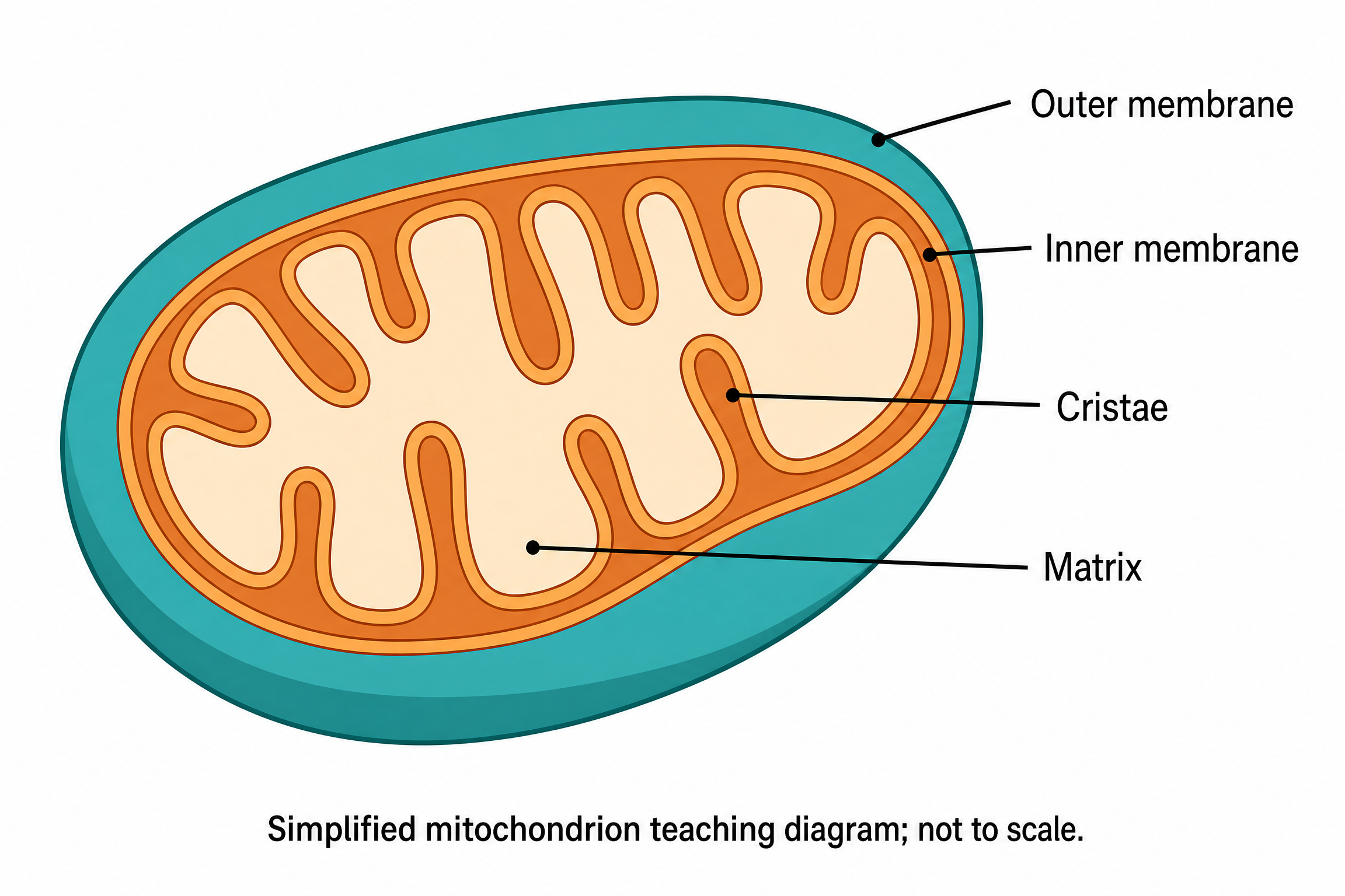
Create a full-color conceptual teaching cutaway of one mitochondrion on white. Show a smooth outer membrane, a distinct inner membrane folding inward into cristae, and matrix enclosed by the inner membrane. Use exactly four labels with non-crossing leader lines: Outer membrane, Inner membrane, Cristae, Matrix. Outer membrane must point to the outside boundary, Inner membrane to the inner boundary, Cristae to one inward fold of the inner membrane, and Matrix to the interior compartment. Use contrasting teal and orange membranes with a pale interior. Do not add ATP, electron transport proteins, reaction arrows, dimensions or measured results. Add the exact footer: Simplified mitochondrion teaching diagram; not to scale.
Reviewed output · self-authored simulated input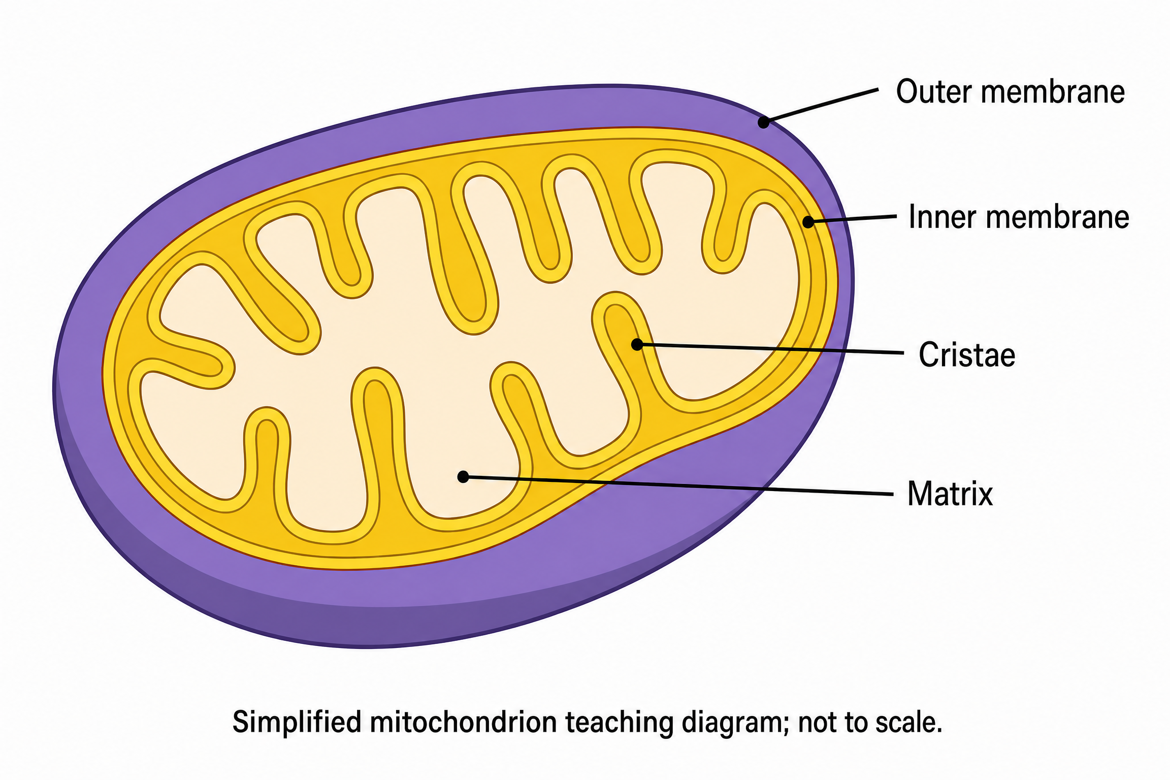
Use a permitted self-authored PNG from the four-label mitochondrion cutaway as a reference. Change the outer membrane to violet and inner membrane to gold. Preserve the smooth outer boundary, connected cristae folds, matrix compartment, all four exact labels and their correct endpoints, and the simplified/not-to-scale footer. Add no reaction arrows or molecular machinery.
Reviewed output · self-authored simulated inputRead the example input
{
  "style": "flat-color",
  "brief": "Create a full-color conceptual teaching cutaway of one mitochondrion on white. Show a smooth outer membrane, a distinct inner membrane folding inward into cristae, and matrix enclosed by the inner membrane. Use exactly four labels with non-crossing leader lines: Outer membrane, Inner membrane, Cristae, Matrix. Outer membrane must point to the outside boundary, Inner membrane to the inner boundary, Cristae to one inward fold of the inner membrane, and Matrix to the interior compartment. Use contrasting teal and orange membranes with a pale interior. Do not add ATP, electron transport proteins, reaction arrows, dimensions or measured results. Add the exact footer: Simplified mitochondrion teaching diagram; not to scale."
}
Load the disclosed simulated illustration brief and review every label, connection and scientific detail. AI results vary between runs.
Distinguish the two membranes and the matrix
A mitochondrion cutaway makes the outer membrane, inner membrane and enclosed matrix visible in one teaching illustration. Cristae are folds of the inner membrane. The default brief uses contrasting membrane colors and four exact labels so each label target can be reviewed independently.
The generator uses Image 2 at 2K to produce a separate raster draft. Check that the cristae remain connected folds of the inner membrane and that Matrix points into the compartment enclosed by it. A visually plausible organelle does not verify membrane geometry, molecular machinery, ATP production or measured cell function.
Follow the membrane boundaries
Trace the smooth outside boundary and the separate folded inner boundary. Reject a drawing whose cristae appear detached or whose folds cannot be followed back to the inner membrane.
Inspect all four label endpoints
Outer membrane must identify the outside boundary, Inner membrane the inner boundary, Cristae an inward fold and Matrix the interior compartment. Non-crossing leader lines help inspection but still require a manual check.
Keep structure separate from biochemical function
An organelle illustration is not an assay of respiration or ATP production. Keep measurements and molecular mechanisms out of this example and retain the simplified/not-to-scale disclosure.
Review reference: OpenStax Biology 2e: eukaryotic cells. Check each generated label and structure against an appropriate subject reference.
How to use this tool
- Describe the subject and the exact relationships to show.
- Choose a style and aspect ratio; optionally add one permitted PNG reference.
- Review the brief and explicitly confirm generation.
- Inspect the result against the supplied scientific details.
- Download the original PNG or revise the brief for a new image.
Specifications and limits
- Route
- Independent Image 2 2K raster illustration; separate from native diagram engines
- Controls
- Scientific brief; line art, flat color or semi-realistic style; supported aspect ratios; optional PNG reference
- Reference
- One PNG up to 5 MiB, 8192 pixels per side and 24 million pixels; only material you may send
- Output
- Original PNG; labels are pixels, without SVG, PPTX or independently editable annotations
- Review
- User scientific review required; no scientific-accuracy or publication-acceptance guarantee
Guest native edits stay in this tab until you explicitly save a confirmed figure. Sign in to keep private works and revision history. Saved native works restore the figure and exports, not the original input form. AI tasks, images and references are private to their owner. Subscriptions and one-time packs are available for AI allowances. Native drawing and exports remain free. Understand export formats.
Frequently asked questions
Why does the tool name say mitochondria but the example shows one?
Mitochondria is the plural term; mitochondrion is singular. The default example requests one simplified mitochondrion so its boundaries and four labels can be inspected clearly.
How are cristae different from the inner membrane?
Cristae are folds of the inner membrane. In the teaching cutaway, the Cristae leader line should identify a fold while Inner membrane identifies the inner boundary. Review the generated geometry and both label endpoints.
Where should the Matrix label point?
It should point to the interior compartment enclosed by the inner membrane, not to a membrane line or the space between the two membranes. The default brief states this target explicitly.
Does the diagram show ATP production or the electron transport chain?
The default example deliberately omits ATP, electron transport proteins and reaction arrows. A molecular-process illustration needs its own supplied details and separate scientific review; this generator does not simulate the process.
Can I change the membrane colors or add a PNG reference?
Yes. Supply the requested colors in the brief and optionally attach one eligible PNG you have permission to send. Colors are visual distinctions, not measured properties, and reference-guided generation still needs review.
Can I download editable membrane and label objects?
This route downloads the original PNG. The membranes, labels and leader lines are pixels; it does not provide editable SVG, PPTX objects or a separate annotation layer. Revise and explicitly regenerate for a new draft.